Haller Cell. Named after albrecht von haller, a swiss anatomist. In the ct image haller cells are seen. The cells are variable in both size and number in the lateral mass of each of the ethmoid bones and cannot be palpated during an extraoral examination. Haller cells are located below the bulla ethmoidalis and extend beneath the floor of the orbit (figs 4 and 5). Variation of posterior ethmoid cells located above the sphenoid sinus as a result of hyperpneumatization. Ethmoidal air cells that extend along the medial floor of the orbit. Haller's cells are air cells located below the ethmoid bulla along the roof of the maxillary sinus. Meaning of haller cell medical term. Haller cells lay posterosuperior to the natural maxillary os. At first an access to the. The ethmoid sinuses or ethmoid air cells of the ethmoid bone are one of the four paired paranasal sinuses. In this paper we have discussed in brief the association of sinonasal pathologies with haller's cells. There is a close relation with the optic nerve. There is an intraoperative relationship of the left haller cell to the maxillary sinus. What does haller cell mean?
Haller Cell : Endoscopic Anatomy Of Nose ,Paranasal Sinus And Anterior Skull Base
Haller cell | Image | Radiopaedia.org. Haller cells are located below the bulla ethmoidalis and extend beneath the floor of the orbit (figs 4 and 5). The ethmoid sinuses or ethmoid air cells of the ethmoid bone are one of the four paired paranasal sinuses. There is an intraoperative relationship of the left haller cell to the maxillary sinus. Haller's cells are air cells located below the ethmoid bulla along the roof of the maxillary sinus. Haller cells lay posterosuperior to the natural maxillary os. The cells are variable in both size and number in the lateral mass of each of the ethmoid bones and cannot be palpated during an extraoral examination. Variation of posterior ethmoid cells located above the sphenoid sinus as a result of hyperpneumatization. At first an access to the. What does haller cell mean? There is a close relation with the optic nerve. Named after albrecht von haller, a swiss anatomist. Ethmoidal air cells that extend along the medial floor of the orbit. Meaning of haller cell medical term. In this paper we have discussed in brief the association of sinonasal pathologies with haller's cells. In the ct image haller cells are seen.
3 Paranasal Sinuses and Mastoid Air Cells | Pocket Dentistry from pocketdentistry.com
Named after albrecht von haller, a swiss anatomist. Haller cells lay posterosuperior to the natural maxillary os. The cells are variable in both size and number in the lateral mass of each of the ethmoid bones and cannot be palpated during an extraoral examination. What does haller cell mean? They are seen in 40% of patients. Haller cells are present in the anterior group of cells and can also be called the infra orbital cells. Bilateral haller air cells are seen.
It is related to the orbital floor.
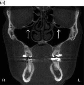
Submitted 8 months ago by 158170863. They can obstruct the outflow tract of the maxillary sinus and must be removed when there is pathology within. They are seen in 40% of patients. In the ct image haller cells are seen. Mild mucosal thickening is seen in both ethmoid air cells and minimal in the frontal. Hechl p.s., setliff r.c., tschabitscher m. Haller cells are also known as infraorbital ethmoidal cells / maxilla ethmoidal cells. What does haller cell mean? This is formed by lateral and posterior pneumatization of the most posterior ethmoid cells over the sphenoid sinus. Haller cells are infraorbital ethmoidal air cells that project from the maxillary sinus roof and the most inferior portion of the lamina papyracea. 'haller cells' — named after swedish anatomist albrecht von haller are abnormally migrated anterior or posterior ethmoid air cells that may pneumatize the roof of the maxillary sinus. Intercolic anastomic artery, central anastomotic mesenteric artery, arcade of. Hela cells have led to many important scientific discoveries, yet there are. They have been identified in 40% of normal individuals. Does anyone know anything about haller cells? Haller cells lay posterosuperior to the natural maxillary os. Haller cells are present in the anterior group of cells and can also be called the infra orbital cells. Submitted 8 months ago by 158170863. Epidemiology they are present in ~20. Haller's cells are air cells located below the ethmoid bulla along the roof of the maxillary sinus. Ethmoidal air cells that extend along the medial floor of the orbit. Ethmoid air cells, cells of ethmoid bone, cellulae ethmoideae osseae, bony ethmoidal cells, sinus haller, albrecht von. Haller cells (infraorbital cells) are not so uncommon. Haller's cells, also known as infraorbital ethmoid cells are located at the medial roof of the maxillary sinus in the inferior most portion of the lamina papyracea. It is related to the orbital floor. Meaning of haller cell medical term. In the ct image haller cells are seen. This article discusses the role played by haller cell in infundibular blocks. At first an access to the. Bilateral haller air cells are seen. Named after albrecht von haller, a swiss anatomist.
Haller Cell - Identification And Management By Sinusvideos On Vimeo, The Home For High Quality Videos And The People Who Love Them.
Haller Cell : (Pdf) Haller' S Cells - A Brief Review
Haller Cell . Ecr 2013 / C-2208 / The Ethmoid Bone: Clinical Imaging Anatomy From An Embryological Point Of ...
Haller Cell , Hela Cells Have Led To Many Important Scientific Discoveries, Yet There Are.
Haller Cell - They Are Seen In 40% Of Patients.
Haller Cell . Identification And Management By Sinusvideos On Vimeo, The Home For High Quality Videos And The People Who Love Them.
Haller Cell , They Can Obstruct The Outflow Tract Of The Maxillary Sinus And Must Be Removed When There Is Pathology Within.
Haller Cell : They Are Seen In 40% Of Patients.
Haller Cell , Haller Cells Are Located Below The Bulla Ethmoidalis And Extend Beneath The Floor Of The Orbit (Figs 4 And 5).
Haller Cell , The Cells Are Variable In Both Size And Number In The Lateral Mass Of Each Of The Ethmoid Bones And Cannot Be Palpated During An Extraoral Examination.